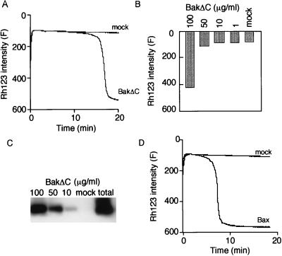
Figure 1.
Δψ loss induced by rBakΔC and rBax in isolated mitochondria. (A and B) Δψ loss induced by rBakΔC. Isolated mitochondria (1 mg/ml) were incubated with 100 μg/ml (A) or the indicated concentration (B) of rBakΔC, and Δψ was measured by using Rh123 uptake over 20 min (A) or at 20 min (B). (C) Uptake of rBakΔC into mitochondria. After the same experiments as in B were performed, mitochondria (30 μg) were centrifuged, washed, and subjected to Western blot analysis using anti-Bak antibody. The total amount of rBakΔC protein in 100 μg/ml is indicated as total. (D) Δψ loss induced by rBax. Isolated mitochondria (1 mg/ml) were incubated with 100 μg/ml rBax, and Δψ was measured by using Rh123 uptake over 20 min.

