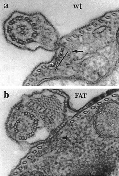
Figure 5.
Transmission electron micrographs of thin sections through the flagellum area in control (a) and 20-hr FAT (b) cells. The flagellum of control cells (wt) contains the axoneme consisting of 9 + 2 microtubules, the PFR, and a filamentous structure (arrow) that extends from the PFR into the cell body, where it interrupts the regular spacing of the cortical microtubules. Four adjacent microtubules (microtubule quartet, indicated by a bar) immediately to the left of the filamentous structure are distinct because of their association with endoplasmic reticulum cisternae and other properties discussed in the text. In at least one flagellum of FAT cells, the axoneme lacks the central microtubule pair and some of the outer doublets are missing or have an altered morphology, whereas the PFR appears normal. Also missing is the microtubule quartet and some cortical microtubules in the area where the flagellum is anchored to the cell body through the filament system (arrow). (×100,000.)

