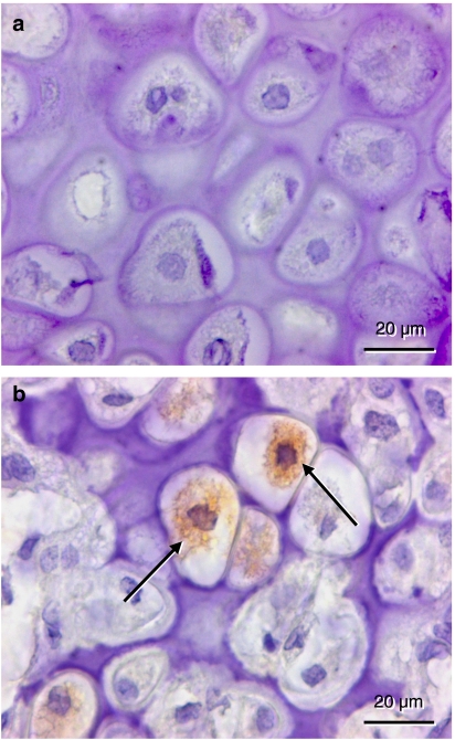
Figure 5.
Immunohistochemistry of vascular endothelial growth factor (VEGF) expression within the callus of a rapamycin-treated (a) and a control animal (b) at 2 weeks post-fracture. In contrast to the rapamycin-treated animal, several hypertrophic chondrocytes of the control animal show positive staining for VEGF in the cytoplasm (arrows).

