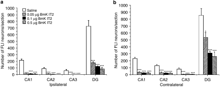
Figure 5.
Histograms of the number of labelled FLI neurons in the ipsilateral (a) and contralateral (b) hippocampus in the saline and BmK IT2 groups. The number of labelled FLI neurons in the hippocampus from 9 to 11 sections of each animal was counted and averaged (n=6–8) 2 h after the injection of pilocarpine. Data are shown as mean±s.e.mean. *P<0.05 and ***P<0.005 compared with the saline group (One-way ANOVA, Tukey's test).

