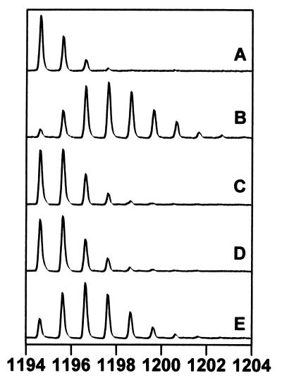
Figure 3.
MALDI-TOF mass spectra of one of the eight peptides from the PKA-PKI(5–24) analysis that experienced slowed exchange in the complex. The spectra are expanded so as to show the isotopic distribution for the ion of interest (MH+ = 1194.6463). (A) The undeuterated peptide. The higher mass peaks in the envelope are caused by naturally occurring isotopes. (B) The isotopic envelope for the same peptide obtained from the PKA sample that was deuterated for 10 min before pepsin digestion. The additional peaks are due to the incorporation of deuterium in the peptide. (C) The peptide obtained from the deuterated PKA sample that was allowed to off-exchange for 10 min. (D) The peptide from the PKA-Mg2+ATP complex that was allowed to off-exchange for 10 min. (E) The peptide from the PKA-Mg2+ATP/PKI(5–24) complex that was allowed to off-exchange for 10 min.

