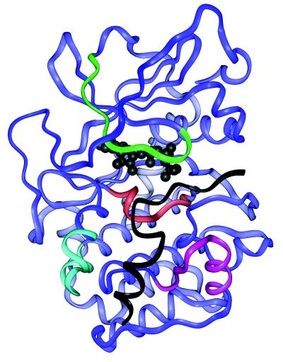
Figure 4.
Structure of the PKA-Mg2+ATP/PKI(5–24) complex [PKA in blue, PKI(5–24) in black, and ATP in ball-and-stick, black] (13) showing the location of sequences that experienced slowed off-exchange in the complex. Three peptides identified the ATP-binding site and catalytic loop (residues 164–172, red). Three peptides identified the glycine-rich loop (residues 44–54, green). One peptide corresponded to the substrate-binding shelf (residues 237–250, purple) and one peptide corresponded to a second contact with the PKI(5–24) (residues 133–145, cyan).

