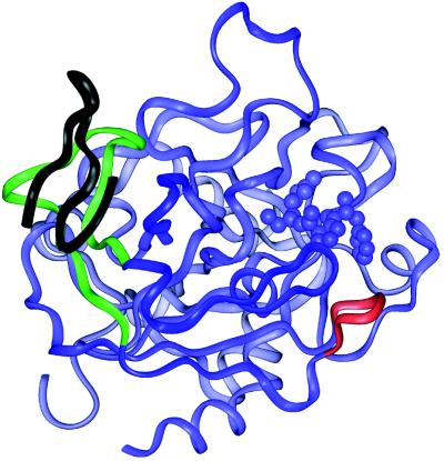
Figure 7.
Structure of human α-thrombin (blue) complexed to a 19-aa residue fragment from TM (black) and to an active site inhibitor (blue) (14) showing two sequences that experienced slowed off-exchange in the complex. The sequence that corresponds to the anion-binding exosite I (residues 97–117) is colored green. An additional sequence of decreased solvent accessibility upon TMEGF(4–5) binding (residues 127–132), is shown in red.

