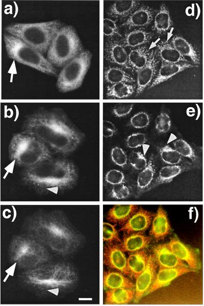
Figure 4.
PAT1 is a cytoplasmic protein enriched in the perinuclear region. (a–c) Paraformaldehyde-fixed HeLa cells were labeled with mAb26 (a and b) and visualized by using the TSA system with HRP-conjugated secondary antibody and Cy3-tyramide. Cells in b are were also double-labeled with anti-tubulin antibody and visualized with fluorescein isothiocyanate-labeled secondary antibody (c). Note that PAT1 gives a punctate cytoplasmic staining, and in some cells the staining is clearly enriched in the perinuclear region (arrows in a and b) which colocalilizes with the microtubule organization center (MTOC) as detected by heavily concentrated microtubules (arrow in c). (Bar = 10 μm.) (d–f) Acetone-fixed HeLa cells were double-labeled with mAb26 (d) and anti-APP antibody (e) and visualized without TSA technique with rhodamin and fluorescein isothiocyanate-labeled secondary antibodies, respectively. The filamentous labeling of PAT1 is more obvious in these cells (arrows), which overlap with APP (arrowhead). The overlap of APP and PAT1 distribution is clearly seen as the orange signal in the merged image (f). The data showing the antibody specificity are presented on the PNAS web site (www.pnas.org) as Fig. 4 g and h.

