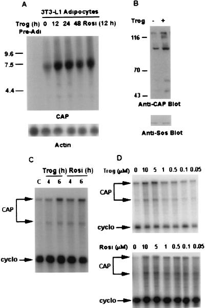
Figure 1.
Activation of CAP gene in 3T3-L1 adipocytes by PPARγ activators. (A) 3T3-L1 adipocytes were treated with troglitazone (10 μM) for the indicated times or with rosiglitazone (10 μM) for 12 h. CAP and β-actin mRNA expressions were measured by Northern blot analysis sequentially. (B) Cell lysates prepared from 3T3-L1 adipocytes treated with troglitazone (10 μM) for 12 h or left untreated were analyzed directly by immunoblotting with anti-CAP antibodies (anti-CAP Blot) or anti-Sos antibodies (Anti-Sos Blot). (C) 3T3-L1 adipocytes were treated either with troglitazone or rosiglitazone (10 μM) for the indicated times or were left untreated. RNase protection assay was performed on total RNA (10 μg/per sample) as described in Materials and Methods. (D) 3T3-L1 adipocytes were treated with the indicated concentrations of either troglitazone or rosiglitazone for 12 h, and 10 μg/sample total RNA was analyzed by RNase protection assay. Trog, troglitazone; Rosi, rosiglitazone; cyclo, cyclophilin.

