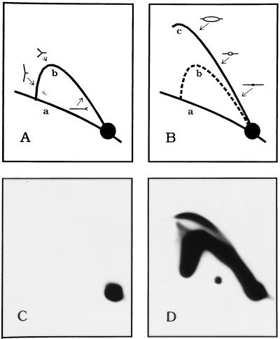
Figure 4.
Neutral/neutral 2-D gel analysis to confirm cell cycle position. (A and B) The 2-D gel replicon mapping method (38) separates a digest of replication intermediates in the first dimension according to molecular mass and in the second dimension according to both mass and shape. The nonreplicating restriction fragments in the genome trace a diagonal of linear fragments (curve a) whereas fragments containing single replication forks (A, curve b) or internal initiation sites (replication bubbles; B, curve c) are separated cleanly from the linear fragments and from each other. By hybridizing a transfer of such a gel with a probe specific for a fragment of interest, its mode of replication can be discerned. (C). 2-D gel analysis of a 6.2-kb EcoRI fragment containing ori-β in DNA isolated from cells blocked at the G1/S boundary with mimosine (see Materials and Methods). No replication intermediates can be detected. (D) Analysis of the same ori-β-containing fragment 90 min after removal of mimosine, when initiation is at the peak in this locus.

