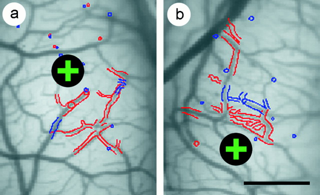
Fig. 1.
Alignment of anatomical sections to optical maps. The alignment of the first two anatomical sections (redand blue, respectively) with an image of the superficial blood vessels is shown for a normally sighted kitten (a) and for a kitten exposed to 2 d of strabismus (b). In both a andb, the injection site of the anatomical tracer is marked with a black circle enclosing a green +. Computer-assisted drawings of the anatomical sections were independently scaled, rotated, and translated to achieve the best alignment with the vascular image. Outlines of the pial vasculature were used to align the first two sections with the vascular image. Coevident profiles of radial blood vessels were used to align deeper sections with more superficial sections. In this manner, the distribution of retrogradely labeled cells and anterogradely labeled boutons were brought into register with the optical maps of ocular dominance. Scale bar, 1 mm.

