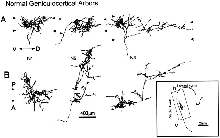
Fig. 3.
Computer reconstructions of PHA-L-immunostained axonal arbors in area 17 in a normal P49 animal. All geniculocortical arbors in normal, OND (Fig. 4), and OD (Fig. 5) animals were obtained from the dorsal-most portion of the medial bank of the lateral gyrus.A shows the arbors as originally reconstructed in the coronal plane, and B shows the arbors as seen from the pial surface, after a 90° rotation along the dorsoventral axis of the lateral gyrus. The arrowheads indicate the boundaries of layer IV. V↔D = ventrodorsal axis indicated inA; A↔P = anteroposterior axis indicated in B. Inset, Drawing of coronal section showing arbor N3 and rectangle in the medial bank of the lateral gyrus containing all arbors reconstructed.Inset: D, Dorsal direction;V, ventral direction.

