Figure 1.
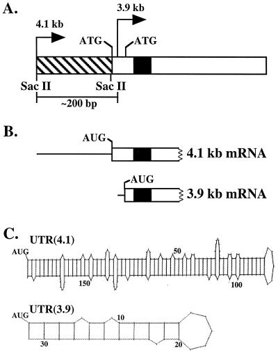
(A) Schematic representation of exon one of the murine β4GalT-I gene. The bent arrows denote the position of the 4.1-kb transcriptional start site (4.1 kb) and the 3.9-kb transcriptional start site (3.9 kb) relative to the two in-frame ATGs that are separated by 39 bp. The 3.9-kb start site is positioned between the two in-frame ATGs, ≈200 bp downstream of the 4.1-kb start site. The hatched and open rectangles represent the 5′-UTR and coding sequence of the 4.1-kb mRNA, respectively. Note the UTR(3.9) is embedded within the coding sequence of the 4.1 kb-mRNA. The black box indicates the position of the transmembrane domain. (B) The 5′ end of the 4.1- and 3.9-kb β4GalT-I mRNA is shown, where the thin line and open rectangle represent the 5′-UTR and part of the coding sequence, respectively. Translation of the 4.1- and 3.9-kb β4GalT-I mRNA results in two catalytically identical, trans-Golgi resident protein isoforms with NH2-terminal cytoplasmic domains of 24 and 11 aa, respectively. (C) Potential secondary structure of the 5′-UTR of the 4.1- and 3.9-kb β4GalT-I transcript. The long UTR(4.1) has a calculated ΔG of −76 kcal/mol, whereas the short UTR(3.9) has a calculated ΔG of only −7 kcal/mol (13).

