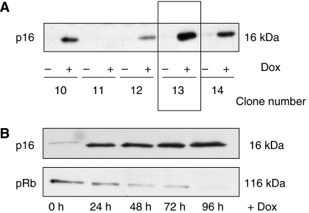
Figure 1.
Generation MiaPaCa-2-TREx–p16 cells with doxycyclin-inducible expression of p16. (A) The plasmids pcDNA6/TR and pcDNA4/TO-p16 were sequentially transfected into p16-deficient MiaPaCa-2 cells and clones with inducible expression of p16 were selected (MiaPaca-2-TREx-p16). For each of the zeozine-resistant MiaPaCa-2-TREx-p16 clone, cells were grown for 48 h in the presence (+Dox) or absence (−Dox) of 1 μg ml−1 doxycycline. The expression of p16 protein was analysed by western blotting. The clones with the highest p16 induction and lowest basal expression were further analysed, for example clone 13. (B) Time course of doxycycline-induced p16 expression. Cells were cultured for 96 h in the presence or absence of doxycycline and each 24 h, lysates were prepared for western blot analysis of p16 (upper panel) and pRB (lower panel). Equal amounts of protein (20 μg) were separated on sodium dodecyl sulphate polyacrylamide gel electrophoresis.

