Figure 1.
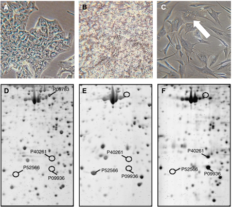
Morphology and proteome pattern differing in hepatocarcinoma, B-lymphoblastoid, and myofibroblastoid cell-lines. Light microscopy of HCC-1.2 in (A), BLC-1 in (B), and MF-2 cells in (C). Arrow in (C) indicates lipid droplets. Magnification: × 80. In (D–F) cytosolic proteins of the lines were separated by 2D-PAGE and detected by fluorography. Selected proteins were further identified by mass spectrometry (Zwickl et al, 2005). A segment of a representative 2D-PAGE gives highly different protein profiles in (D) HCC-1.2, in (E) BLC-2, and in (F) MF-2 cells. Swiss Prot numbers: P05783, keratin type 1, cytoskeletal 18; P09936, ubiquitin carboxyl-terminal hydrolase isozyme L1; P40261, nicotinamide N-methyltransferase; and P52566, rho GDP-dissociation inhibitor 2.

