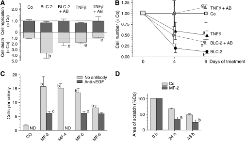
Figure 4.
B-lymphoblastoid cells induce death of hepatocarcinoma cells whereas myofibroblastoid cells enhance neoangiogenesis and migration of hepatocarcinoma cells. In (A) and (B): HCC-2 cells were treated 24 and 72 h after seeding and were harvested after 4 and 6 days. Abbreviations of treatment groups: (Co), untreated HCC-2; (TNFβ), aliquots of a TNFβ stock (Sigma-Aldrich; 1 μg ml−1 PBS/0.1% BSA) were added for finally 1.5 ng ml−1 medium; (BLC-2), HCC-2 exposed to medium supernatant conditioned by BLC-2; (TNFβ+AB) or (BLC-2+AB), TNFβ-containing medium or conditioned supernatant were pre-incubated with anti-TNFβ. In (A), cells were kept for 96 h. 3H-thymidine was added 24 h before harvesting, and DNA replication was determined by autoradiography. To assay apoptosis by FACS-analyses, cells were incubated in 0.5 ml PBS containing 15 μg propidium iodide (Sigma-Aldrich) for 30 min at 4°C and were analysed in a Becton-Dickinson FACSCalibur system. In (B) cells were harvested and counted. In (A) and (B) means±s.d. from three separate experiments are given. Statistics by Kruskal–Wallis test; Co vs cell supernatant or TNFβ: (a) P<0.05; (b) P<0.01; cell supernatant vs neutralised supernatant or TNFβ vs neutralised TNFβ: (c) P<0.05; (d) P<0.01. In (C) HUVEC were seeded at 1 × 10³ per cm2. After cell attachment supplements in M199-medium were reduced to 1% FCS and no ECGS for 24 h before start of treatment. Abbreviations of treatment groups: (Co), control medium; (MF-2), (MF-4), (MF-5), or (MF-6), medium supernatant conditioned by the MF-cells. Control media or conditioned supernatants were pre-incubated with anti-vEGF. Treatments were renewed after 72 h for further 96 h. The size of the HUVEC colonies was determined by counting the number of cells. Experiments were performed in triplicate and at least 10 colonies per well were scored. Abbreviations: ND, not done. In (D) confluent HCC-2 cultures were scratched manually with a 200 μl pipette tip, followed by rinsing and treatments. Abbreviations of treatment groups: (Co), control medium; (MF-2), medium supernatant conditioned by MF-2 cells. Total area of the scratches was measured by morphometry (Lucia 6.0, Nikon, Düsseldorf, FRG). In (C) and (D) mean±s.e.m. of at least three independent studies are given. Statistics by Kruskal–Wallis test; Co vs cell supernatant: (a) P<0.05; (b) P<0.01; cell supernatant vs neutralised supernatant: (c) P<0.05.

