
Figure 2.
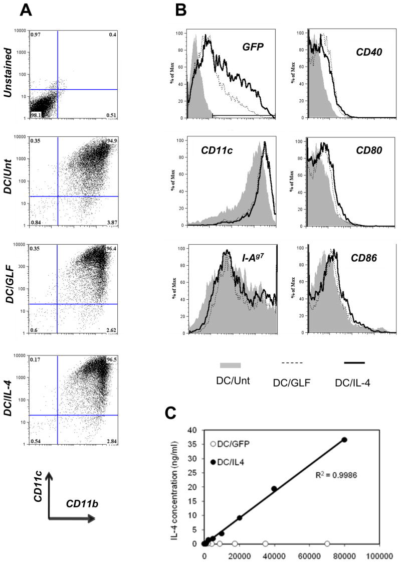
Characterization of DCs used for adoptive cellular gene therapy. Untransduced DCs (DC/Unt) and transduced DCs (DC/GLF and DC/IL-4) were analyzed by flow cytometry. Plots were all gated on live DCs based on FSc and PI profile. (A) Purity was determined based on CD11c and CD11b expression. (B) Transduction efficiency was assessed by GFP expression on live DCs. Surface marker levels (CD11c, CD40, CD80, CD86 and class II MHC I-Ag7) were analyzed on total live DCs for DC/Unt (grey-filled histograms), or on the GFP+ live DC fraction for DC/GLF (dashed line) and DC/IL-4 (thick line). (C) After harvest on day 6, transduced DCs (DC/GFP and DC/IL-4) were re-plated at various cell numbers in cytokine-free medium for another 36h, after which supernatant was collected and used for ELISA (one of two representative experiments shown).