
Figure 5.
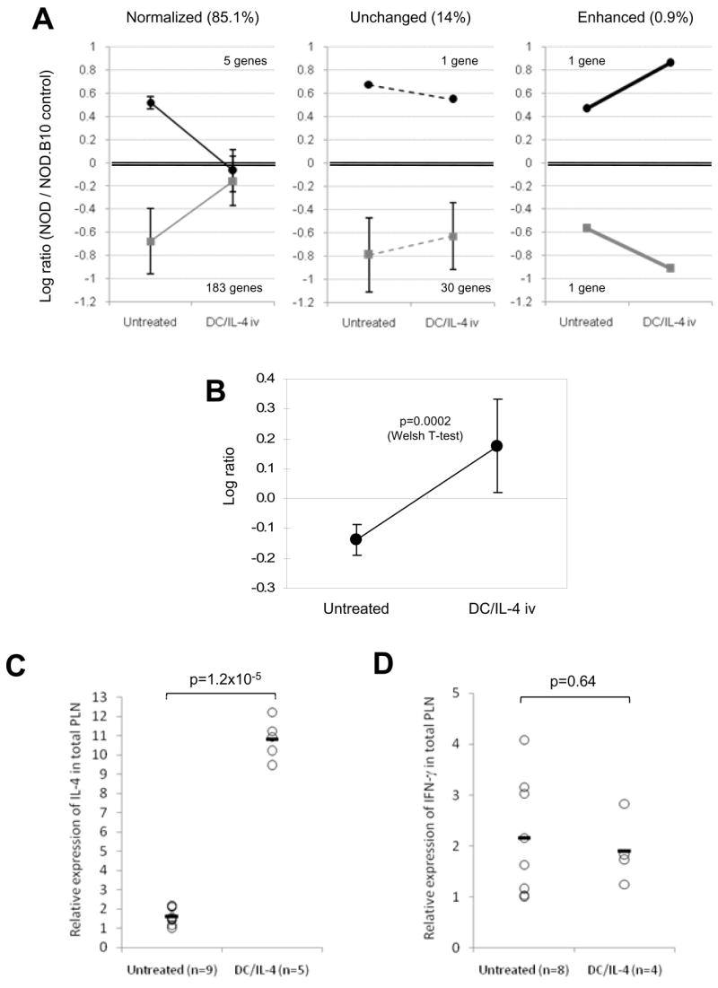
Changes in gene expression induced by DC/IL-4 in the PLN of 12 wk-old NOD mice. (A) Gene expression in PLN from female NOD mice, untreated (n=7) or treated with DC/IL-4 iv (n=9), was analyzed against a tissue-matched control from female NOD.B10 mice. A total of 221 genes, which expression was ~3 fold over- (log10 ratio >0.45) or under-expressed (log10 ratio <-0.45) in NOD compared to NOD.B10 (p<0.01) were selected. Their change, 3 days following treatment with DC/IL-4, was graphically represented in three groups (normalized, unchanged or enhanced; mean of log10 ratios ± SD) using data shown on Table 1. Normalized or enhanced gene expression were significantly changed by DC/IL-4 treatment (p<0.05). (B) IL-4 expression in NOD PLN, relative to NOD.B10 control, before and after DC/IL-4 treatment (mean ± SD; p=0.0002, T-test). IL-4 was not part of the above genes, because although significantly under-expressed, its log10 ratio was <0.45. (C, D) Relative expression of IL-4 (C) and IFN-γ (D) by RT-PCR in PLN of untreated or DC/IL-4-treated 12-wk old NOD mice (individual results and mean; data normalized to β actin; T-test applied).