Figure 4.
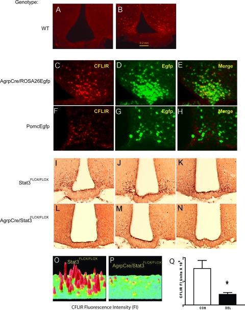
Fasting-induced rise in CFLIR was decreased in Agrp/Npy neurons in DEL mice. CFLIR by immunofluorescence in the arcuate nucleus in the fed state (A) and after 48 h fasting (B). Fasting-induced CFLIR (C) and green fluorescent cells expressing Egfp (D) in AgrpCre/ROSA26Egfp mice are shown. E, Colocalization of CFLIR and Egfp in AgrpCre/ROSA26Egfp mice. Fasting-induced CFLIR (F) and green fluorescent cells (G) expressing Egfp in PomcEgfp mice. H, CFLIR and Egfp in PomcEgfp mice. I, CFLIR by avidin biotin complex staining in the arcuate of 48-h fasted 8- to 12-wk-old mice was greater in CON mice (I–K) than DEL littermates (L–N). O and P, Surface plots of CFLIR by immunofluorescence after 48 h fasting in CON (O) and DEL littermates (P). Q, Means ± sem of summed FI units for fasting-induced CFLIR by immunofluorescence in arcuate nucleus of CON and DEL littermates (n = 4/group).

