Figure 1.
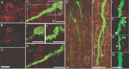
GnRH neuron dendrites express Na+v channels. A, Projected confocal image of Na+v-ir in the CA1 hippocampus of GnRH-GFP mice; B, single optical slice (0.36 μm) through two neurons (asterisks) showing with membrane-localized Na+v-ir; C, omitting Na+v primary antibody eliminates Na+v-ir; D, projected confocal stack of a GnRH soma and proximal dendrite (green) and Na+v-ir (red): i–iii, below, single optical slices (0.36 μm) corresponding to regions indicated by white boxes in D; E, low-power projected confocal image of GnRH neuron cell bodies and dendrites and Na+v-ir; F, projected confocal stack of a portion of dendrite 390 μm from the cell body of a GnRH neuron shown in E (white box); i–iii, far right, single optical slices (0.36 μm) corresponding to regions indicated by white boxes in F, with arrows highlighting yellow pixels indicative of Na+v-ir localized in the distal segment of a GnRH dendrite; G, omitting Na+v primary antibody eliminates Na+v-ir. Scale bars, 10 μm (A–E) and 5 μm (G).

