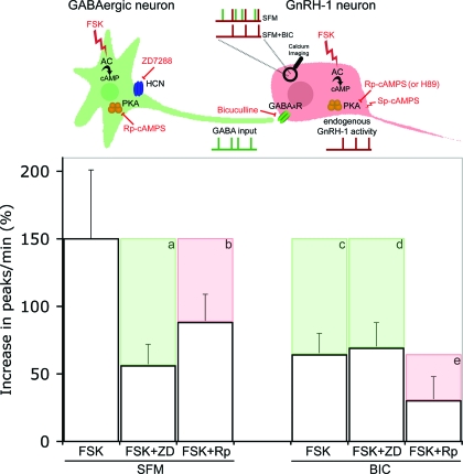
Figure 9.
Summarized data. The upper panel represents the cellular partners evaluated in this study: GnRH-1 neurons (red) with their GABAergic input (green). Independent endogenous electrical activity of GnRH-1 and GABAergic neurons are represented by, respectively, red and green ticked lines under cells. The resulting intracellular calcium oscillations are recorded using calcium imaging, symbolized by a magnifying glass on the GnRH-1 cell body, and are represented by ticked lines above the GnRH-1 cell. Two experimental conditions are represented: SFM, which allows one to record the summation of the two cells’ endogenous activity (green/red), and SFM+BIC, which allows one to record the endogenous GnRH-1 neuronal activity (red). Pharmacological actions are symbolized in red. FSK stimulates AC, ZD7288 (ZD) blocks HCN channels, BIC antagonizes GABAA receptors, Rp-cAMPS (Rp) and H89 block PKA, and Sp-cAMPS stimulates PKA. Histograms show the percent increase in the number of peaks per minute in response to FSK under different experimental conditions. Pharmacological inhibitions are represented by shaded boxes showing loss of response (a–e). When GnRH-1 neurons are driven by GABAergic input (SFM), ZD inhibits the FSK-induced stimulation (a), whereas Rp is not as effective (b). However, in the presence of BIC, ZD fails to block the FSK-induced stimulation (d) in contrast with Rp (e). The inhibition induced by BIC alone (c) reflects the GABAergic contribution to the FSK-induced stimulation (c, green). Note that the inhibition of the FSK response induced by ZD in SFM (a) is similar to that detected after removal of GABAergic input alone (c) and in the presence of BIC+ZD (d). This suggests that in SFM, the inhibition exerted by ZD occurs on HCN channels localized to GABAergic neurons (a, green). The inhibition induced by Rp in BIC (e) can be directly attributed to a PKA-dependent pathway in GnRH-1 neurons (e, red) and likely corresponds to the inhibition observed in SFM (b, red), even though the PKA pathway cannot be totally excluded in GABAergic neurons.

