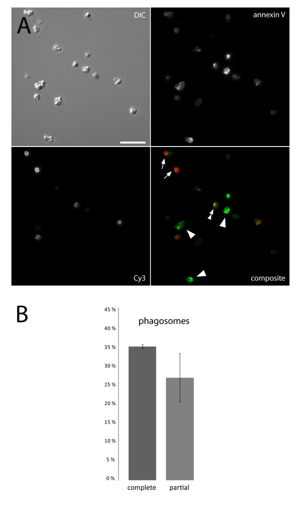
Figure 4.
Phagosome integrity. In A, discrimination of free bacteria from intact and broken phagosomes is illustrated. Alexa 488-labeled annexin V (green) stains phagosomal membrane and Cy3-labeled anti-human Fab fragments (red) stains damaged phagosomes and free opsonized bacteria. Arrows indicate free bacteria, arrowheads intact phagosomes, and the double arrowhead shows a partial phagosome; scale bar 10 μm. B shows quantification of phagosomes from three separate experiments ± SEM.

