
Figure 1. Migration of the embryonic body-wall muscle cells in C. elegans.
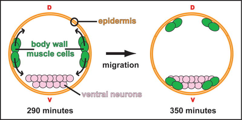
A cross-sectional schematic depicting the migrations of the body-wall muscle cells during embryogenesis. The muscle cells are born as two clusters on either side of the embryo. At 290 minutes (25°C; approximate bean stage) the cell clusters separate at the lateral axis and migrate towards both the dorsal (d) and ventral (v) midlines. The migrating cells pass between the overlying epidermal sheet and the underling cells, which include neuronal precursors. By 350 minutes (25°C; approximate late comma stage) the cells have taken up positions flanking what will become the dorsal and ventral nerve cords (Hresko et al., 1994).