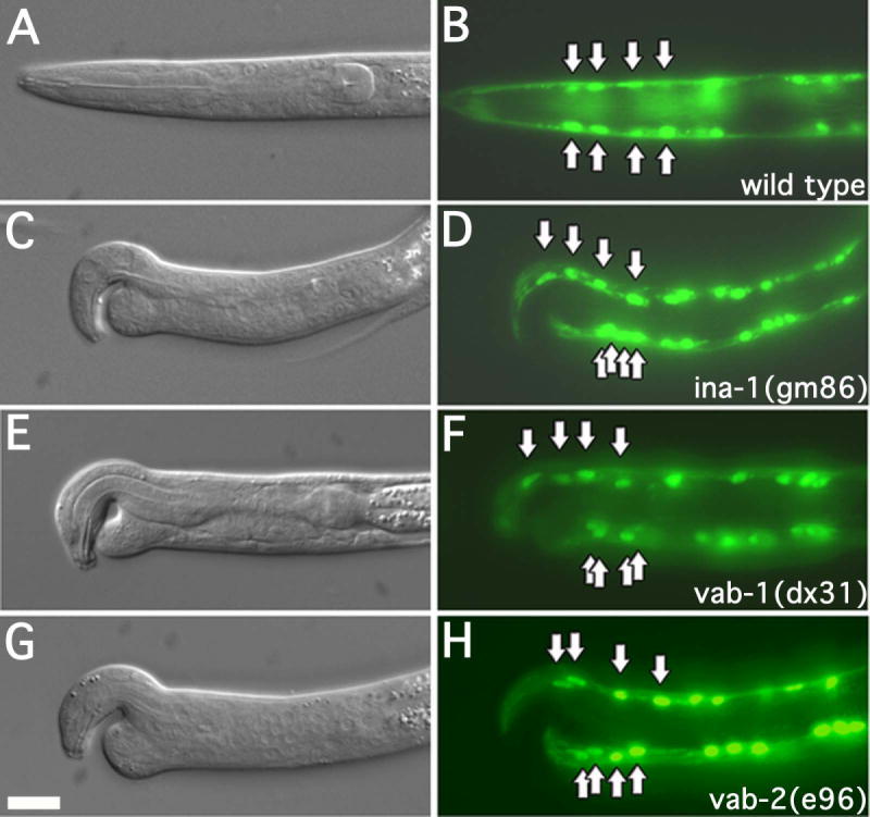
Figure 2. Muscle cells are mispositioned in notched head worms.

DIC and fluorescence images of L1 larva expressing myo-3::GFP. Arrows indicate position of the anterior most muscle cell nuclei. The genotypes of the strains are as indicated. Every case in which larva displayed an apparent notched head phenotype, the anterior most ventral muscle cell nuclei were mispositioned. More than 50 L1 larva were examined for each strain. In this and all subsequent figures dorsal is to the top, anterior is to the left. Scale bar represent 10μm.
