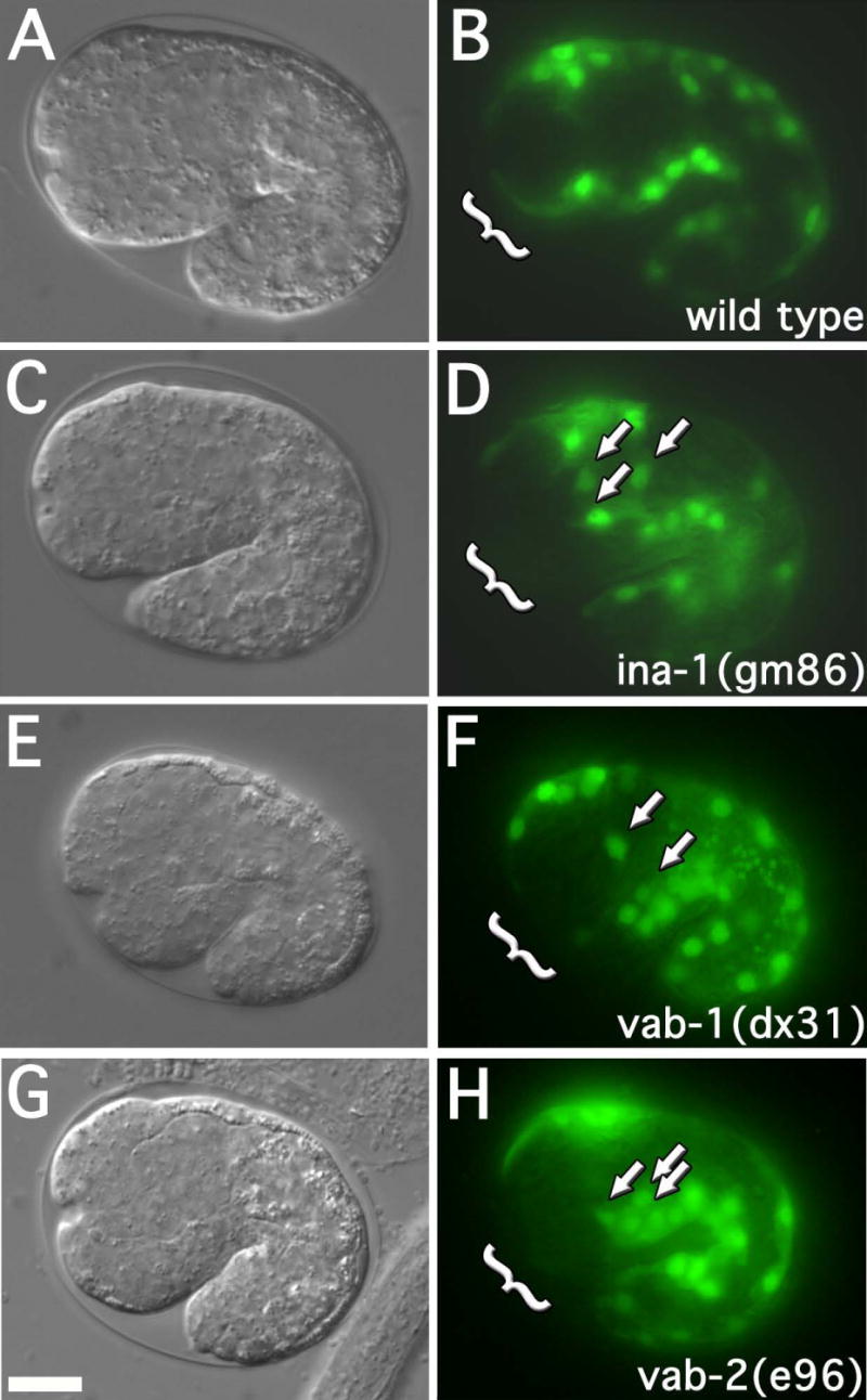
Figure 3. Muscle cell migration defects in ina-1, vab-1 and vab-2 mutant worms.

DIC and fluorescence images of approximate 1½ fold embryos (25°C; approximately 380 minutes) expressing hlh-1::GFP. The genotypes of strains are as indicated. Arrows indicate mispositioned muscle cell nuclei. Brackets indicate the anterior-ventral quadrant. Embryos at this stage were assayed for anterior muscle cell nuclei misplaced towards the posterior or lateral surface. 30 embryos were examined for each strain. The frequency of misplaced muscle cell nuclei were as follows: wild type (2 of 30, 7%), ina-1(gm86) (21 of 30, 70%, p<0.001), vab-1(dx31) (23 of 30, 77%, p<0.001), and vab-2/efn-1(e96) (18 of 30, 60%, p<0.001). p-values represent comparison to wild type. Scale bar represent 10μm.
