
Figure 4. Notched head and muscle cell migration defects in mnp-1(RNAi) worms.
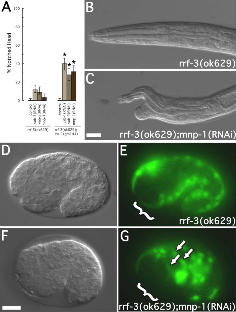
(A) Notched head RNAi phenotype. Strains include rrf-3(ok629) and ina-1(gm144);rrf-3(ok629). RNAi treatment included vab-1(RNAi), vab-2(RNAi), and mnp-1(RNAi). For each treatment, 200 L1 larval were examined. Asterisks indicate a significant difference (p<0.0002) between rrf-3(ok629) and ina-1(gm144);rrf-3(ok629) for a given RNAi treatment. Error bars represent 95% confidence intervals. (B-C) DIC images of L1 larvae showing the mnp-1(RNAi) notched head phenotype. Genotypes and RNAi treatment are as indicated. Scale bar represent 10μm. (D-G) DIC and fluorescence images of approximate 1½ fold embryos expressing hlh-1::GFP. Arrows indicate mispositioned muscle cell nuclei. Brackets indicate the anterior-ventral quadrant. Embryos at this stage were assayed for anterior muscle cell nuclei misplaced towards the posterior or lateral surface. The frequency of misplaced muscle cell nuclei were as follows: rrf-3(ok629) (7 of 129, 5.4%) and rrf-3(ok629)::mnp-1(RNAi) (39 of 339, 11.5%, p<0.05). Scale bars represent 10μm.