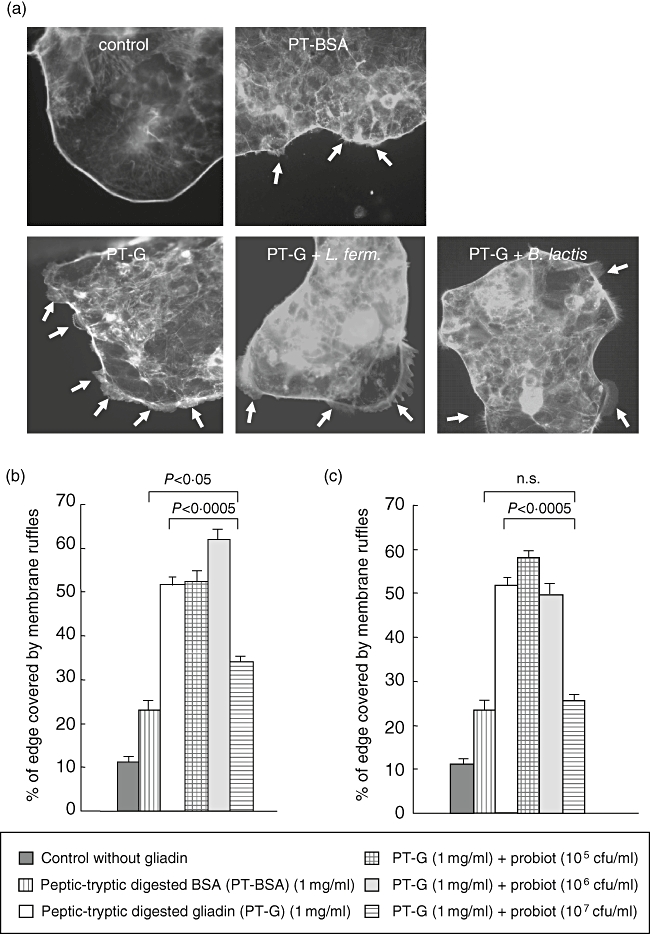
Fig. 2.

Membrane ruffle formation in Caco-2 cells in the presence of gliadin and the two different bacterial strains. (a) Caco-2 cells cultured without gliadin show a uniform cell cluster edge without any membrane protrusions while PT-BSA induced some ruffle formation. Caco-2 cell clusters which have received gliadin have large membrane ruffles on their edge. The addition of 107 colony-forming units (cfu)/ml of both Lactobacillus fermentum and Bifidobacterium lactis together with gliadin reduced the formation of the ruffles. Arrows point to membrane ruffles. (b) Addition of L. fermentum to Caco-2 cells cultures supplemented with gliadin at a lower concentration does not inhibit the membrane ruffle formation induced by gliadin. At a concentration of 107 cfu/ml L. fermentum reduced membrane ruffle formation from 51·6% to 34·1%. (c) The lowest concentrations of B. lactis do not protect from gliadin-induced membrane ruffle formation, while B. lactis at a concentration of 107 cfu/ml is able to lower the percentage of cellular edge covered by ruffles from 51·5% to 25·7%. The bars represent data measured from five pictures in duplicate samples performed three times. The error bars represent standard error of the mean. Different experimental settings are indicated in the box below. P-value < 0·05 is considered significant. PT-BSA, peptic-tryptic-digested bovine serum albumin; PT-G, peptic-tryptic-digested gliadin.
