
Fig. 3.
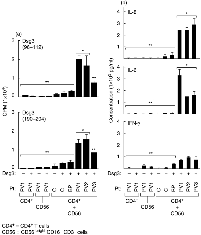
Specific presentation of desmoglein 3 (Dsg3) peptides to CD4+ T cells obtained from skin lesions of pemphigus vulgaris (PV) patients by CD56+ major histocompatibility complex (MHC) class II-positive natural killer (NK) cells. CD4+ T cells were obtained from perilesional skin of three untreated PV patients with active disease and interacted with CD56+ MHC cells II-positive NK cells of the same patient, in the presence of two different Dsg3 peptides. (a) Dsg3 (a.a. 96–112) and Dsg3 (a.a. 190–204) were used. T cell proliferation was statistically significantly higher in CD56+ NK cells compared with controls (P < 0·02). Responses to Dsg3 (96–112) were statistically significantly higher than the response to Dsg3 (190–204) (P < 0·03). (b) Supernatants collected from these cultures are presented. Levels of cytokines produced in response to both Dsg3 peptides were similar. The levels of interleukin (IL)-8 are statistically significantly higher than levels of IL-6 (P < 0·02). Interferon-γ levels were also increased but not statistically significant. Controls included CD4+ T cells from skin biopsies of two normal controls and one patient with bullous pemphigoid. Bars represent the standard deviation. *P < 0·05; **statistically not significant.