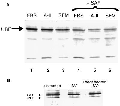
Figure 2.
TBP Far Western blot analysis of SMC nuclear extracts showing enhanced TBP–UBF binding in growth-stimulated SMC that was reduced by treatment with SAP. (A) Far Western blot of TBP binding proteins in SMC nuclear extracts. Cells were growth-arrested in SFM and then stimulated with A-II or FBS as described in Methods. Nuclear extracts were prepared as described (23). Four μg of each extract were treated with either SAP (10 units/ml, United States Biochemical) or glycerol (vehicle) and were incubated for 1 h at 37°C before electrophoresis and Far Western blotting. Lanes 1–3 show blots of nuclear extracts from SMC treated with 10% FBS, A-II, or SFM vehicle, respectively. Lanes 4–6 show blots of nuclear extracts treated with SAP. The band corresponding to the UBF1/UBF2 doublet is indicated, although the two UBF isoforms are not well resolved under the conditions of these assays. (B) Control Western blot analyses with a UBF antibody confirmed the position of UBF and demonstrated that treatment with SAP had no effect on UBF protein content. Similar results to these were obtained when TBP Far Western analyses were performed by using immunoprecipitates of UBF derived from vehicle and growth factor-stimulated SMC (data not shown), as compared with the whole nuclear extracts as shown here.

