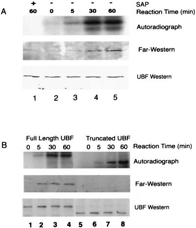
Figure 3.
Far Western analysis of rUBF1 phosphorylated in vitro showed phosphorylation-dependent UBF–TBP binding. (A) 32P-autoradiograph analysis (Top) of rUBF1 phosphorylated in vitro by SMC nuclear extract. The phosphorylation reaction was performed as described in Methods. SAP was added for the last 30 min of one of the 60-min time point samples. The membrane was dried and exposed to film for 18 h to detect 32P incorporation into rUBF1. Far Western analysis (Middle) of TBP binding to rUBF1 was performed as described in Methods. Control UBF Western blot (Bottom) showing equal amounts of rUBF in each of the experimental groups. (B) Autoradiographic analysis of 32P incorporation (Top) or Far Western blot analysis of TBP binding (Middle) by using truncated rUBF lacking the C-terminal acidic tail (amino acid residues 1–656, a generous gift from L. Rothblum, Weis Research Institute, Danville, PA) or full-length rUBF. (Bottom) A UBF Western blot showing equal amounts of rUBF in each of the experimental groups. Phosphorylation reactions and Far Western analyses were performed as described in Methods, with incubation times as indicated. Western blot analyses with UBF antibody showed that equivalent amounts of truncated and full-length rUBF1 were present in each lane (data not shown).

