
Fig. 3.
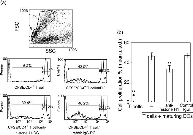
Histogram plots show the cell division associated with carboxyfluorescein succinimidyl ester (CFSE)-labelling of Piebald Virol Glaxo CD4+ T cells stimulated by mature dendritic cells (DCs) alone or by a DC culture in the presence of either anti-histone H1 antibody or control immunoglobulin G (IgG). (a) All plots were gated on CD4-positive cells (histograms not shown), while the histograms were also gated to include both resting lymphocytes (R1) and blasts (R2). CFSE-labelling of CD4+ T cells alone or T cells stimulated by mature DCs, DCs treated with anti-histone H1 antibody or DCs treated with control IgG. (b) A histone H1 blockade induced by anti-histone H1 antibody abrogates T cell proliferation. Data are representative of three independent experiments with essentially similar tendencies. **P < 0·01.