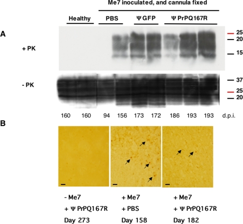
Figure 3. Analysis of PrPSc accumulation in the brains of mice after treatment with lentiviral vectors.
Immunoblot analysis of brain homogenates before and after proteinase K digestion (A). Control mice were either not inoculated with prions or were inoculated with Me7. Mice inoculated with the Me7 prion strain and implanted with a guide cannula received injections of either PBS, lentivirus carrying the GFP gene, or lentivirus carrying the PrPQ167R gene. The numbers below indicate the dates when the mice were killed in days post-inoculation with prions (d.p.i). The numbers to the right of each blot indicate the molecular mass of the protein standards in kDa. A mix of anti-PrP antibodies was used to stain blots of SDS-polyacrylamide gels. Immunohistochemical detection of PrPSc deposits in the thalamus sections (B). The plus (+) and minus (−) signs indicate that the mice were or were not inoculated with the ME7 prion strain and whether they were injected with PBS or PrPQ167R virions. Tissue labelling was performed on 9 animals (3 per group). Pictures are representative of the staining observed in different animals in each group. Arrows indicate the localization of the PrPSc deposits in the tissue sections. Bar, 6 µm.

