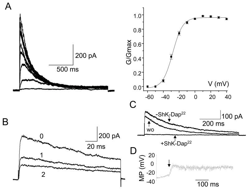
Figure 2. Electrophysiological and pharmacological properties of Kv1.3 channels in SLE T cells.
A. Kv1.3 currents were recorded in whole-cell configuration and were elicited by depolarizing voltage steps from −60 mV to +40 mV (10 mV increments) from −80 mV HP every 30 sec. The conductance-voltage curve (constructed from current amplitudes such as those shown) was fitted to a Boltzman function and the voltage at which half of the channels are activated (V1/2) calculated. B. Kv1.3 channels’ cumulative inactivation was induced by consecutive depolarizing pulses applied every second. The maximal current amplitude progressively decreased with each successive pulse (indicated by progressive numbers). C. Effect of Shk-Dap22 on Kv1.3 current in SLE T cells. Currents were recorded in whole cell configuration in physiologic solution before application of Shk-Dap22 (-ShK-Dap22), after Shk-Dap22 (10 nM) inhibition and after drug wash-out (wo). D. Membrane potential (MP) measured by current-clamp before and after ShK-Dap22 (10 nM) addition. The time of ShK-Dap22 introduction is indicated by an arrow. The SLEDAI of the patients for this study ranged from 2–12.

