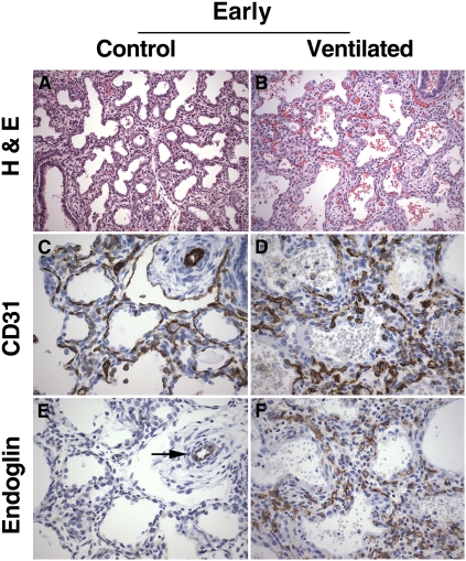
Figure 3.
Lung histology and platelet endothelial cell adhesion molecule (PECAM)-1/endoglin immunohistochemistry of early control subjects and short-term ventilated patients. (A) Early control lung showing large-sized, simple airspaces, relatively wide septa, and focal early secondary crest formation, characteristic of late canalicular/early saccular stage of lung development (infant born at 24.5 wk of gestation, lived for 30 min). (B) Short-term ventilated lung showing widening and increased cellularity of the septa, associated with focal hemorrhages within the air spaces (infant born at 23 wk, lived for 5 d, ventilated). (C) PECAM-1 (CD31) immunostaining of early control lung, highlighting the double subepithelial capillary network characteristic of the canalicular/saccular stage of lung development. (D) PECAM-1 (CD31) immunostaining of short-term ventilated lung, showing abundant, disarrayed capillary structures within the expanded septa. (E) Endoglin immunostaining of early control lung corresponding to (C), showing endoglin immunoreactivity in association with a peripheral arteriole (arrow). Endoglin staining is virtually absent in capillary structures within the septa. (F) Endoglin immunostaining of short-term ventilated lung corresponding to (D), showing abundant endoglin-positive capillary structures within the widened septa. (A and B) Hematoxylin–eosin staining (H&E); (C and D) PECAM-1 (CD31) immunohistochemistry, DAB (3,3′-diaminobenzidine tetrachloride) with hematoxylin counterstain; (E and F) endoglin (CD105) immunohistochemistry, DAB with hematoxylin counterstain. Original magnification: (A and B) ×200; (C–F) ×400.

