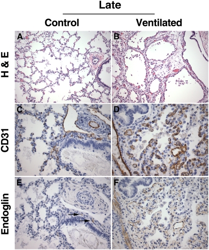
Figure 4.
Lung histology and platelet endothelial cell adhesion molecule (PECAM)-1/endoglin immunohistochemistry of late control subjects and long-term ventilated patients. (A) Late control lung showing late saccular/early alveolar architecture characterized by thin alveolar septa and abundant secondary crests (nonmacerated stillborn delivered at 36 weeks gestation, cause of death unknown). (B) Long-term ventilated lung showing simple, large-sized airspaces with wide and hypercellular septa (infant born at 24 weeks, lived for 56 days, ventilated). (C) PECAM-1 (CD31) immunostaining of late control lung, showing branching capillary structures within the alveolar septa and immunoreactive endothelium in central arterial and lymphatic structures. (D) PECAM-1 (CD31) immunostaining of long-term ventilated lung, showing abundant, primitive-appearing and predominantly nonbranching capillary structures within the expanded septa. (E) Endoglin immunostaining of late control lung corresponding to (C), showing weak endoglin immunoreactivity limited to rare central capillary structures (arrows). Endoglin staining is absent in the interstitial microvasculature. (F) Endoglin immunostaining of long-term ventilated lung corresponding to (D), showing diffuse endoglin immunoreactivity in association with capillary structures in the widened septa. (A and B) Hematoxylin–eosin staining (H&E); (C and D) PECAM-1 (CD31) immunohistochemistry, DAB (3,3′-diaminobenzidine tetrachloride) with hematoxylin counterstain; (E and F) endoglin (CD105) immunohistochemistry, DAB with hematoxylin counterstain. Original magnification: (A and B) ×200; (C–F) ×400.

