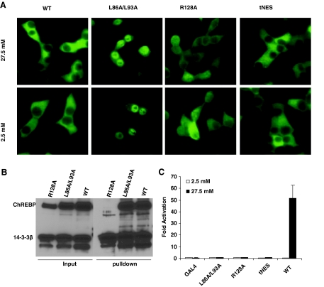
Figure 2.
Subcellular Localization and Glucose Response of ChREBP and Its Mutants
A, Immunofluorescent staining. The 832/13 cells stably expressing c-myc-tagged wild-type or indicated mutant ChREBP were treated with low (2.5 mm) or high (27.5 mm) glucose for 12 h before staining with anti-c-myc antibody. B, Poly-His pull-down assay. pCHM-14-3-3β was cotransfected with plasmids expressing indicated wild-type or mutant ChREBP into 832/13 cells. Twenty-four hours after transfection, we used Western blot with anti-c-myc antibody to examine the levels of expressed proteins in the cell lysate (input) or in the precipitate after pull-down assay. C, Luciferase assay. pG5-luc and pRL-TK were cotransfected with indicated plasmids into 832/13 cells that were treated with 2.5 or 27.5 mm glucose for 24 h. WT, Wild type.

