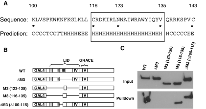
Figure 6.
Identification of the 14-3-3-Interacting Motif in ChREBP
A, Secondary structure prediction with SSpro8 program. Key residues were marked by asterisks and their number in the ChREBP amino acid sequence. The conserved α-helix (116–135) is boxed. Codes for secondary structure: C, coil; H, α-helix; T, turn; E, extended strand. B, Schematic illustration of the ChREBP deletion constructs (not to scale). C, Poly-His pull-down assay. pCHM-14-3-3β was cotransfected with indicated plasmids shown in B into 832/13 cells. Cells were lysed 24 h after transfection for poly-His pull-down assay.

