
Figure 2. Immunostaining of 4-HNE and 3-NT increases with age.
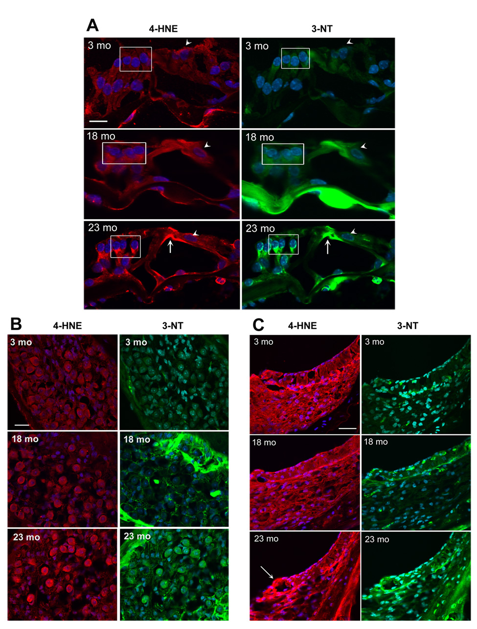
Sections from the basal turn of the cochlea were prepared and stained as described in ‘Methods’. Red fluorescence, 4-HNE; green fluorescence, 3-NT; blue fluorescence, DAPI staining for nuclei. Scale bar: 10 µm.
Panel A: Immunostaining of 4-HNE and 3-NT in the organ of Corti is associated with the phalangeal processes of Deiters cells (box) and the inner and outer pillar cells (arrow). The staining was consistently stronger at 18 and 23 months than at 3 and 12 months (12 months not shown). Staining of inner hair cells was less affected by age (arrow heads). The figures are representative of three animals for each condition.
Panel B: Immunostaining of 4-HNE and 3-NT increased in spiral ganglion cells at the age of 23 months, but not at 18 months. The figures are representative of three animals for each condition. The bright green areas in the 18- and 23-mo sections are bony structures around the ganglion cells.
Panel C: Immunostaining of 4-HNE and 3-NT in stria vascularis increased in specific segments of the lateral wall tissues at the age of 23 months. Mostly affected was the area of the spiral prominence (arrow). At 23 months, the thickness of spiral ligament (fibroblasts and other cell types) appeared reduced. The figures are representative of three animals for each condition.