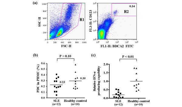
Figure 2.
Proportion of pDCs and IFN-α production capacity. (a) A gating strategy was used to distinguish plasmacytoid dendritic cells (pDCs) from among peripheral blood mononuclear cells (PBMCs). Blood cells were analyzed using flow cytometry. Total PBMCs were gated at R1 and then analyzed for the presence of blood dendritic cell antigen (BDCA)-2 and CD123. Both BDCA-2-positive and CD123-positive cells were identified as pDCs in gate R2. (b) The proportion of pDCs among PBMCs in patients with systemic lupus erythematosus (SLE) versus healthy control individuals. The solid bars represent the mean percentage of pDCs among PBMCs (0.23% in SLE patients versus 0.30% in controls; P > 0.10). Statistical significance was analyzed using the Mann-Whitney U-test. (c) Relative IFN-α producing capacity in SLE patients versus healthy control individuals. CpG ODN2216-induced IFN-α production was divided by the absolute number of pDCs. (d) Expression of Tll-like receptor (TLR)9 mRNA. TLR-9 expression in PBMCs was measured using semiquantitative reverse transcription PCR. The expression of TLR9 is presented relative to β-actin expression. Each analysis was performed in triplicate, and the average values are indicated by a solid square for SLE patients and solid triangle for healthy control individuals. The solid bars represent the mean value for each experimental group. Statistical significance was analyzed using the Mann-Whitney U-test.

