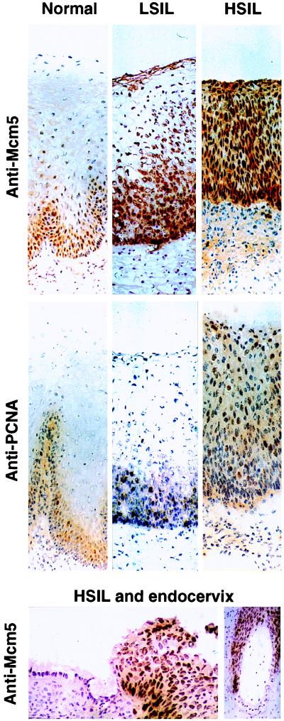
Figure 2.
Immunoperoxidase staining of frozen sections of normal cervix, LSILs, and HSILs with antibodies against PCNA and Mcm5. Frozen sections of normal cervix, LSILs, and HSILs were immunostained for Mcm5 or PCNA (as a conventional proliferation marker) by the immunoperoxidase method. In normal cervix, surface cells likely to be sampled by cervical smear examination are negative with both antibodies. In LSIL with associated koilocytosis (cells showing HPV cytopathic effect), anti-PCNA antibodies stain basal and some parabasal nuclei, but surface koilocytes are negative. In contrast, anti-Mcm5 antibodies stain nuclei in superficial as well as basal epithelial layers including most koilocytes. In HSIL, anti-PCNA antibodies show focal staining of 10% of nuclei in the surface layers, whereas anti-Mcm5 antibodies stain virtually all nuclei, including those at the epithelial surface. Antibodies against Ki-67 (another conventional proliferation marker), show essentially similar results to anti-PCNA. In contrast, antibodies against Cdc6 stain all cells in HSIL as seen with anti-Mcm5 (data not shown; Top and Middle; magnification ×108). Although HSIL cells show strong nuclear immunostaining with anti-Mcm5, endocervical surface, and glandular cells are negative (Bottom Left, magnification ×225; Bottom Right, magnification ×87).

