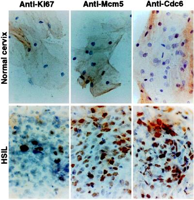
Figure 3.
Immunoperoxidase staining of cells obtained by cervical smear examination from surface of normal ectocervix or HSIL with antibodies against Ki-67, Mcm5, or Cdc6. Superficial squamous cells from normal ectocervix show no nuclear staining with any of the antibodies tested. In HSIL, anti-Ki-67 antibodies (and anti-PCNA; data not shown) show nuclear staining of only a minority population of the abnormal cells in the exfoliated sheets. In contrast, anti-Mcm5 and anti-Cdc6 antibodies stain most HSIL cells. Magnification: ×240.

