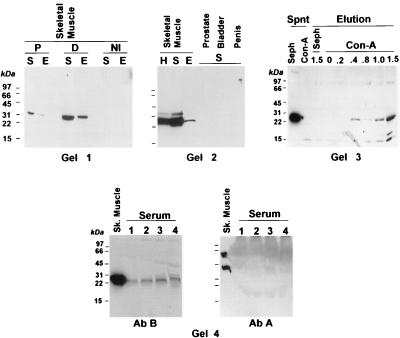
Figure 4.
Western blot of myostatin-related protein in the skeletal muscle and human serum. Tissue extracts were prepared from mouse (Gel 1) and human skeletal muscle (Gel 2) and fractionated into supernatant (S) and membrane extracts (E). H, unfractionated homogenate. (Gel 1) The Western blot membranes were cut into three sections and reacted with either nonimmune IgG (NI) or antibody B directly (D), or in the presence of an excess of the corresponding peptide (P). (Gel 2) The S fraction also was prepared from human prostate, bladder, or penile corpora cavernosa. (Gel 3) The S fraction from the human skeletal muscle was incubated with either Con A-Sepharose (Con-A) or Sepharose (Seph). After centrifugation, both the supernatant (Spnt) and the eluates with increasing concentrations of α-methyl-d-mannoside (Elution) were submitted to Western blot assays with antibody B. (Gel 4, Left) The S fraction of the human skeletal muscle and increasing amounts of human serum (lane 1, 0.2 μl; lane 2, 0.5 μl; lane 3, 1 μl, lane 4, 2 μl) were subjected to Western blot assays with antibody B. (Gel 4, Right) The blot from Gel 4, Left was stripped and reacted with antibody A.

