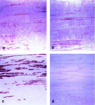
Figure 5.
Immunohistochemical staining. (a) Sections of skeletal muscle were incubated with antibody B (×50). (b) Higher magnification (×100) of a. (c) Serial sections were stained with myosin antibody to identify type I and II fibers. (d) Negative control with antibody B preadsorbed with 7 μg/liter of peptide B.

