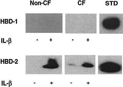
Figure 3.
Detection of HBD-1 and HBD-2 proteins in non-CF and CF ASL by acid/urea/PAGE and immunoblotting. Epithelia were cultured with (+) and without (−) stimulation. (Upper) Comparison of HBD-1 peptide abundance in non-CF and CF epithelia. No HBD-1 peptide was found in washings from non-CF or CF epithelia, but 5 ng of recombinant HBD-1 peptide (STD) was easily detected. No HBD-1 was detected in the cell lysates (not shown). (Lower) Comparison of HBD-2 abundance in non-CF and CF epithelia. The apical surface was washed with water to remove accumulated peptide prior to IL-1β treatment. HBD-2 peptide was easily detected in the washings from non-CF and CF epithelia. Little peptide was recovered with the subsequent NH4OAc wash, indicating good recovery of protein with aqueous washes (not shown). HBD-2 was also detected in the cell lysate (not shown). With IL-1β stimulation, protein recovery increased in all fractions. Results for CF cells are qualitatively similar to those from cultured non-CF airway epithelia. Control was 7.5 ng of HBD-2. Representative results are shown.

