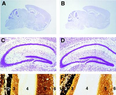
Figure 1.
Brain anatomy of 5-HT2C receptor mutant mice. No phenotypic differences were detected in cytoarchitecture. Cresyl violet staining of sagittal brain sections from wild-type (A) and hemizygous mutant (B) mice. Cresyl violet staining of hippocampal coronal sections from wild-type (C) and mutant (D) mice. Timm’s staining of hippocampal coronal sections revealed no differences in the laminar organization of terminal fields between wild-type (E) and mutant (F) animals. The layers correspond to 1, dentate gyrus granule cells; 2, dentate inner molecular layer; 3, dentate mid/outer molecular layer; 4, stratum lacunosum-moleculare; 5, stratum radiatum; and 6, CA1 pyramidal cells.

