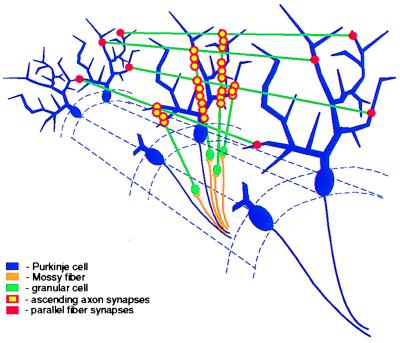
Figure 5.
A schematic diagram of the components in the cerebellar cortex studied here, with Purkinje cells in blue and granular cells in green. The red–yellow circle marks the synapses formed by the ascending axon on Purkinje-cell dendrites, and the red circle marks the synapses formed by the parallel fibers on Purkinje cells along their path. The inhibitory network is not shown.

