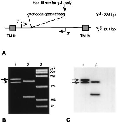
Figure 1.
Scheme illustrating primers used to amplify and to verify the identity of γ2S and γ2L mRNAs. (A) The amplified region of γ2S and γ2L occurred between the third and fourth transmembrane regions (TM III and TM IV), with the kinase-added 5′ forward primer indicated by ∗. (B) Ethidium bromide-stained polyacrylamide gel of amplified γ2S and γ2L cDNAs from a control brain at the predicted molecular weights of 201 and 225 bp (arrows in lane 1). Lane 2, same products digested with HaeIII, isolating the γ2L cDNA product. Lane 3, HaeIII-digested pBS to show molecular-weight markers. (C) Film autoradiogram of same gel showing the uncut, forward-primed 201- and 225-bp radiolabeled products in lane 1 (arrows) and forward-primed 201-bp fragment of γ2S (upper band) and the HaeIII-digested 108-bp fragment of γ2L in lane 2.

