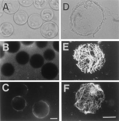
Figure 1.
Formation of nascent β-glucan fibrils on protoplasts. (A) Protoplasts of tobacco cells. (B) Freshly prepared protoplasts at the onset of incubation in 0.01% Calcofluor solution. (C) Calcofluor staining of nascent β-glucan regenerated on protoplasts after 30 min of incubation. (D) A phase-contrast image of the plasma membrane sheet. (E) An immunofluorescence micrograph of cortical microtubules on the membrane sheet. (F) Calcofluor staining of the microfibrils of nascent β-glucans. (Magnification bar is 12 μm.)

