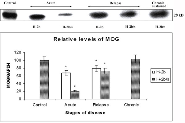
Figure 2.

Distinct patterns of MOG reduction between H-2b/s and H-2b mice throughout disease. Distinct patterns of MOG-specific demyelination were observed between H-2b and H-2b/s mice throughout EAE. Relative levels of MOG were measured similarly as levels of MAG (Fig.1). As opposed to regulation of MAG, most profound depletion of MOG was observed in H-2b/s mice during acute EAE. Lesser then in H-2b/s, loss of MOG was noted in acute H-2b mice during acute disease. Loss of MOG, lesser then in acute but significant compared to control values, was equally found in H-2b and H-2b/s mice with relapsing disease. In H-2b/s mice with chronic-sustained disease levels of MOG were comparable to control values. Stripped and reprobed membrane was shown. Corresponding levels of GAPDH are shown in Fig. 6.
