Figure 7.

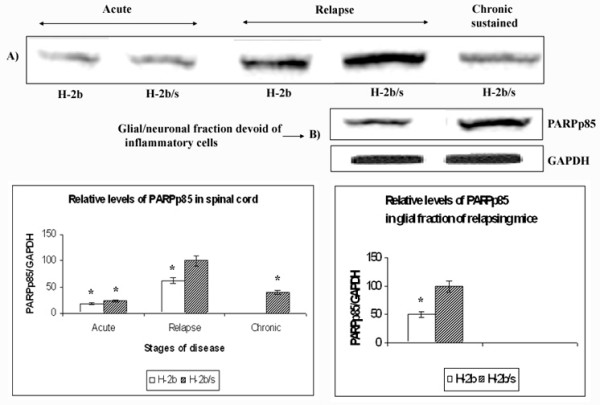
Differences in levels of PARPp85 between H-2b/s and H-2b EAE mice. Relative levels of PARPp85 were measured in spinal cord (A) and in Percol gradient separated fraction of spinal cord, which was devoid of infiltrating inflammatory cells (B). Relative levels of PARPp85 in spinal cord were determined similarly as levels of MAG (Fig. 1). The highest level of PARPp85 was found in relapsing H-2b/s mice, and this value was arbitrarily taken as 100. Compared to this highest level, significantly lower relative levels of PARPp85 were observed during acute and chronic disease. Stripped and re-probed membrane was shown for levels of PARPp85 in spinal cord. Corresponding levels of GAPDH are shown in Fig. 6.
